Anomalia Peters mascată de sechelele unui traumatism ocular
Alina Nagherneac¹, Flavia Chioveanu², Oana Georgiana Maxim³, Daniela Andronic⁴, Alisa Bejan⁵, Anisia Iuliana Alexa⁶
Spitalul Clinic Judeţean de Urgenţă ”Sf. Spiridon’’ Iaşi, România
Cuvinte cheie: anomalia Peters, traumatism ocular, glaucom secundar
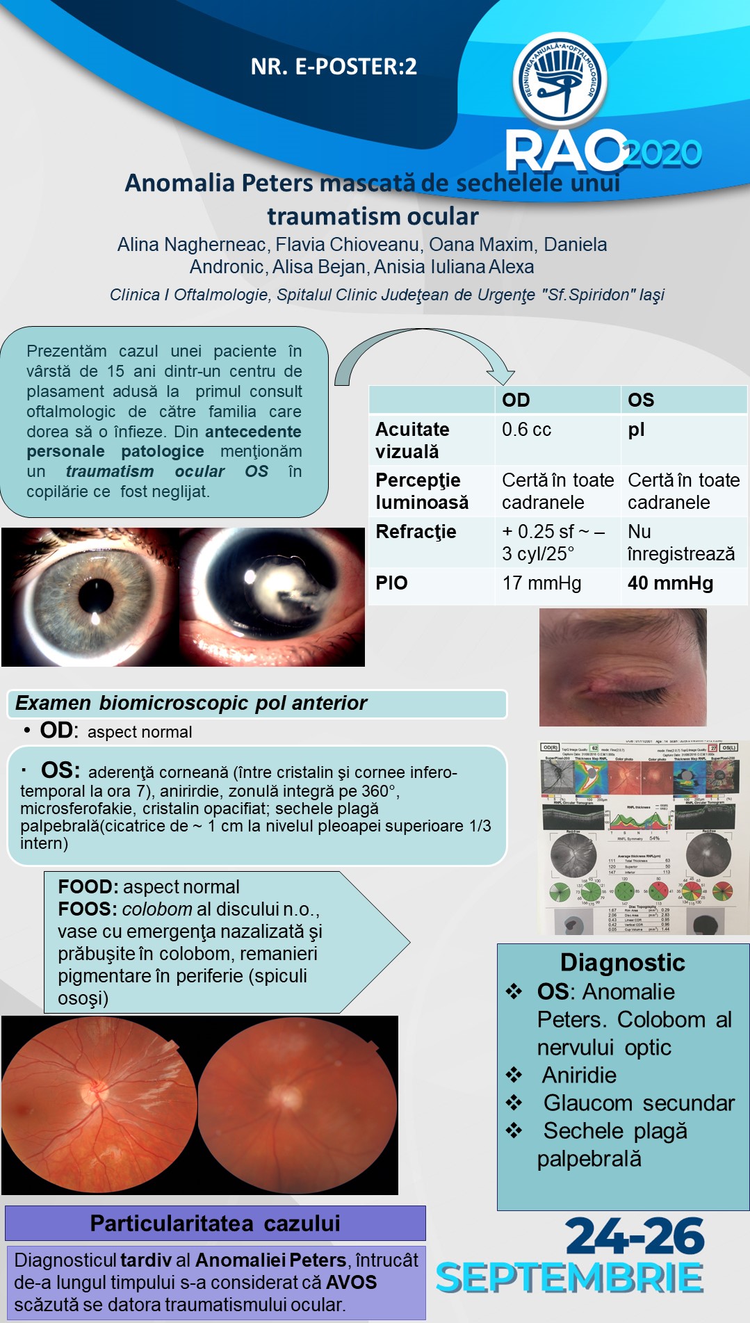
Prezentăm cazul unei paciente în vârstă de 15 ani dintr-un centru de plasament adusă la primul consult oftalmologic de către familia care dorea să o înfieze. La prima vedere se observă la nivelul pleoapei superioare OS o cicatrice, iar pacienta declară în antecedente un traumatism ocular din copilărie ce a fost neglijat, motiv pentru care familia solicită evaluarea globului ocular stâng în vederea stabilirii sechelelor traumatismului ocular. Examenul clinic relevă AVOD 0.6 cc şi PIOOD 17 mmHg, iar la OS o AV scăzută (pl cert) şi PIOOS 40 mmHg, la examenul biomicroscopic al polului anterior OD în limite normale, iar la OS se distinge o aderenţă corneană între cristalin şi cornee, aniridie, microsferofakie şi cristalin opacifiat, modificări sugestive pentru Anomalia Peters. Examenul FOOD fără modificări patologice, iar la OS se observă colobom al nervului optic, vase cu emergenţa nazalizată şi prăbuşite în colobom, remanieri pigmentare în periferie (spiculi osoşi). OCT OS stratul fibrelor nervoase retiniene peripapilare subţiat în sectorul superior şi temporal. Particularitatea cazului constă în diagnosticul tardiv al Anomaliei Peters, întrucât asistentele sociale ce o îngrijeau pe pacientă au considerat de-a lungul timpului că AVOS scăzută se datora traumatismului ocular.
Peters anomaly masked by the sequelae of eye trauma
A.F. Nagherneac¹, F. Chioveanu², O.G. Maxim³, D. Andronic⁴, A. Bejan⁵, A.I. Alexa⁶
“St. Spiridon” Emergency Hospital, Iasi, Romania
Keywords: Peters anomaly, eye trauma, secondary glaucoma
We present the case of a 15-year old patient from an orphanage brought to the first ophthalmological consultation by the family who wanted to adopt her. At first sight, a scar was observed on the upper eyelid of the left eye, and the patient declares a history of neglected eye trauma in childhood, which is why the family requests the evaluation of the left eye in order to establish the sequelae of eye trauma. Right eye (RE) corrected distance visual acuity was 0.6 log MAR with IOP 17 mmHG, whereas left eye (LE) was light perception and IOP 40 mmHg. Slit-lamp examination of anterior segment of RE within normal limits while in the LE a corneal adhesion between the lens and cornea, aniridia, microspherophakia and opacified lens was observed-suggestive changes for Peters Anomaly. The examination of the fundus of the right eye was within normal limits, whilst in the LE a coloboma of the optic nerve with nasalized emergence of vessels and collapsed in coloboma, bone-spicule pigmentary changes in the periphery were described. OCT examination of optic disc showed thinned peripapillary retinal nerve fibre layer in upper and temporal quadrant. The particularity of the case consists in the late diagnosis of Peters Anomaly, as the social workers who cared for the patient considered over time that low LE visual acuity was due to eye trauma.

